Introducing ViSOAR
 As data acquisition advances, and data sizes increase, the need for tools to process and visualize the results in an effective and efficient manner is becoming increasingly important. The reliance on supercomputers for scientific visualization and analysis is already proving to be a hindrance for wide accessibility to researchers and scientists dealing with large data.
The Scientific Computing and Imaging (SCI) Institute and the Center for Extreme Data Management, Analysis, and Visualization (CEDMAV), in collaboration with ARUP Laboratories and the University of Utah, Department of Neurobiology and Anatomy, have developed ViSOAR--a multi platform visualization application for accessing and processing very large imaging data.
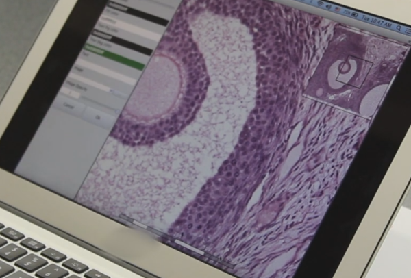
The impetus for the collaboration was to build a tool for the classroom setting that would replace the traditional use of microscopes for viewing histology slides and provide the ability for annotation on the image. As class sizes increase and scanning devices improve in quality, it has become impractical to rely on microscopes for teaching. An additional goal was to make the process mobile – allowing a physician or researcher the ability to review and make diagnoses from anywhere, as opposed to the confines of the lab.
A single scan of a slide can produce multi-gigapixel images that require tens of gigabites of raw data -- thus the need for a tool that can process this data is essential. ViSOAR builds upon the ViSUS technology which allows for large scale data to be streamed over a network, off of a disc, or on the cloud with extreme efficiency. Furthermore, the technology behind ViSUS allows the application to run on a variety of platforms--from mobile devices to desktops to large, multi-display walls. With this approach doctors are liberated from the physical constraints of traditional professional and educational environments and provide remote access to their highly qualified expertise for diagnosing diseases and training the next generation of physicians.
